schedule OP Timing: 8AM to 7PM Monday to Saturday | Emergency Services after 7PM and Sunday

The neuro-ophthalmology department provides expert care for vision problems caused by neurological conditions, bridging the gap between eye health and brain function. We diagnose complex disorders such as optic neuritis, double vision, visual field loss, and cranial nerve palsies—using multimodal imaging and a multidisciplinary approach to protect both sight and neurological well-being.

  • Perimetry in Stroke
  • Non arteritic AION
  • Idiopathic intracranial hypertension
  • Optic Neuritis

Perimetry in Stroke

Perimetry in stroke refers to the assessment of visual field defects in individuals who have suffered a stroke. A stroke can damage the brain's visual processing centers, leading to visual field defects, which can affect a person's daily life and independence.

Perimetry in Stroke

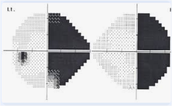
Types of visual field defects in stroke:

  • Hemianopia: loss of vision on one side of the visual field
  • Quadrantanopia: loss of vision in one quarter of the visual field
  • Scotoma: a localized area of vision loss

Perimetry tests used to assess visual field defects in stroke:

  • Confrontation visual field test: a simple test to assess visual field defects
  • Automated perimetry: uses a computerized program to assess the visual field
  • Goldman kinetic perimetry: uses a moving stimulus to assess the visual field
  • Humphrey visual field analyzer: a computerized test to assess the visual field .

Importance of perimetry in stroke:

  • Diagnosis: helps diagnose visual field defects and monitor progression
  • Rehabilitation: guides rehabilitation strategies to improve visual function
  • Safety: identifies potential safety hazards, such as falls or collisions
  • Quality of life: helps individuals adapt to visual field defects and maintain independence

Treatment options for visual field defects in stroke:

  • Vision therapy: rehabilitation strategies to improve visual function
  • Compensatory techniques: teaching individuals to adapt to visual field defects
  • Prisms: using prisms to expand the visual field
  • Assistive technology: using technology, such as magnifying glasses or smart canes, to aid navigation.

Non arteritic AION

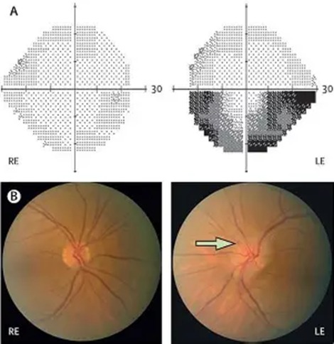
Non-arteritic anterior ischemic optic neuropathy (NAION) is a condition where the blood flow to the optic nerve is suddenly interrupted, causing damage to the nerve and potentially leading to permanent vision loss.

Non arteritic AION

Causes:

  • Spontaneous: exact cause unknown, but thought to be related to a sudden drop in blood pressure or a vascular event.
  • Risk factors: hypertension, diabetes, high cholesterol, sleep apnea, and smoking.

Symptoms:

  • Sudden, painless vision loss in one eye
  • Blurred vision or blind spots
  • Loss of color vision
  • Swelling of the optic disc

Diagnosis is made by clinical examination, visual field testing, optical coherence tomography (OCT) and fluorescein angiography

Treatment:

  • No proven treatment to reverse vision loss
  • Corticosteroids: may be used to reduce swelling and inflammation
  • Aspirin: may be used to prevent further vascular events
  • Lifestyle modifications: controlling risk factors, such as hypertension and diabetes

Prognosis:

  • Variable: some patients may experience partial recovery, while others may have permanent vision loss
  • Recurrence: risk of NAION in the fellow eye is around 15-20%

It's essential to seek medical attention immediately if symptoms occur, as prompt treatment may help reduce the risk of further vision loss.

Idiopathic intracranial hypertension

Idiopathic intracranial hypertension (IIH), also known as pseudotumor cerebri, is a condition characterized by increased pressure within the skull without any apparent cause. The condition is often associated with obesity, and women of childbearing age are more commonly affected.
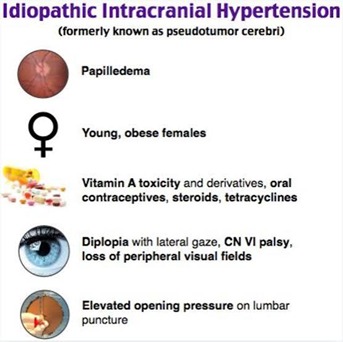
Idiopathic intracranial hypertension

Symptoms:

  • Headaches: often severe and worsening with time
  • Vision problems: blurred vision, double vision, or loss of peripheral vision
  • Papilledema: swelling of the optic disc
  • Tinnitus: ringing or other sounds in the ears
  • Dizziness or vertigo

Diagnosis:

  • Clinical examination
  • Imaging studies (e.g., MRI, CT scan) to rule out other conditions
  • Lumbar puncture to measure cerebrospinal fluid (CSF) pressure
  • Visual field testing to assess vision

Treatment:

  • Weight loss: recommended for obese patients
  • Medications: Acetazolamide to reduce CSF production, Topiramate to reduce CSF production and alleviate symptoms, Corticosteroids to reduce inflammation
  • Lumbar puncture: repeated procedures to drain excess CSF
  • Surgery: Shunting procedures to divert excess CSF ,Optic nerve sheath fenestration to relieve pressure on the optic nerve

Complications:

  • Vision loss: if left untreated or poorly managed
  • Chronic headaches
  • Tinnitus
  • Cognitive impairment

It's essential to seek medical attention if symptoms persist or worsen over time. Prompt treatment can help alleviate symptoms, prevent complications, and improve quality of life.

Optic Neuritis

Optic neuritis is an inflammatory condition that affects the optic nerve, which carries visual information from the eye to the brain. It can cause vision loss, pain, and other symptoms.
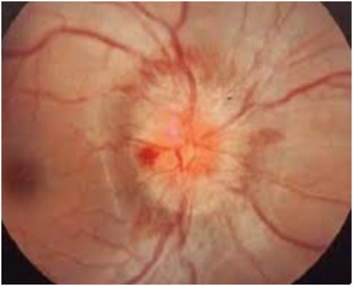
Optic Neuritis

Symptoms:

  • Vision loss: blurred vision, blind spots, or complete vision loss
  • Eye pain: sharp or dull pain, worsened by eye movement
  • Color vision loss: reduced color perception
  • Contrast sensitivity loss: difficulty seeing differences in brightness
  • Afferent pupillary defect: abnormal pupil reaction to light

Causes:

  • Autoimmune disorders: multiple sclerosis, lupus, and rheumatoid arthritis
  • Infections: viral, bacterial, or fungal
  • Inflammatory conditions: sarcoidosis, syphilis

Diagnosis:

  • Clinical examination
  • Visual field testing
  • Optical coherence tomography (OCT)
  • MRI or CT scan to rule out other conditions
  • Blood tests to check for autoimmune disorders or infections

Treatment:

  • Corticosteroids: oral or intravenous to reduce inflammation
  • Pain management: medication to alleviate eye pain
  • Antibiotics or antiviral medication: if caused by an infection
  • Disease-modifying therapies: for underlying autoimmune disorders

Prognosis:

  • Variable: some patients recover fully, while others experience persistent vision loss
  • Recurrence: risk of recurrence, especially in patients with underlying autoimmune disorders

It's essential to seek medical attention promptly if symptoms occur, as timely treatment can improve outcomes and reduce the risk of complications.

Doctors

Dr. Revati Ramesh

DO, FICO

stethoscope_arrow Neuro Ophthalmology, General Ophthalmology

event_available OP Days:

Monday,Tuesday,Wednesday,Thursday,Friday,Saturday